Immuno-fluorescence Microscope
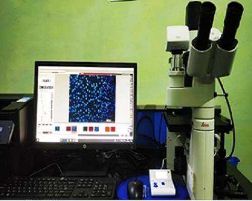
Immunofluorescence (IF) microscopy is a widely used example of immunostaining and is a form of immunohistochemistry based on the use of fluorophores to visualize the location of bound antibodies. It is a particularly robust and broadly applicable method generally used by researchers to assess both the localization and endogenous expression levels of proteins of interest.
Immunofluorescence can be used on tissue sections, cultured cells, or individual cells that are fixed by a variety of methods. Antibodies can be used in this method to analyze the distribution of proteins, glycoproteins, and other antigen targets, including small biological and non-biological molecules.